Scanning Electron and Atomic Force Microscopies for Imaging Protein and RNA Complexes
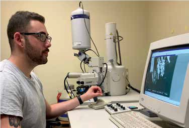
For my capstone project this semester, I have been working on imaging a particular protein and RNA complex using scanning electron microscopy (SEM) and atomic force microscopy (AFM). These methods, SEM and AFM, are utilized in order to visualize extremely small structures all the way from the microscale down to the molecular level. The protein, B-cell activating factor receptor (BAFF-R), and a particular RNA molecule bind to act as a drug delivery system for the treatment of Non-Hodgkin’s Lymphoma. The images collected with these microscopes allow the elucidation of the binding mechanism between the protein and RNA molecules. What’s very exciting about this study, is that this methodology may prove to be a viable means for further studies of protein and RNA drug delivery systems.
This project has been very rewarding because it has allowed me to learn how to operate these very sophisticated scientific instruments and how to analyze the data that they produce. Another aspect that has been particularly valuable is that I am a double-major in physics and chemistry. I have had many struggles with the project throughout the semester, but the knowledge I was able to obtain is extremely valuable. After graduation, I will be starting graduate school at UC Davis for my PhD in chemistry and am very excited to see what unfolds on my next academic journey!